BLOG

Imaging biomarkers play a crucial role in the development, approval, and use of disease-modifying drugs for Alzheimer’s disease

With Alzheimer’s disease (AD) contributing to 60%-70% of the 55 million people living with dementia worldwide, the quest for solutions that slow, or even stop, the underlying disease processes has been a long, sometimes arduous, journey. Beyond the traditional lifestyle recommendations such as physical activity, a healthy weight, and avoiding smoking, people at risk of AD have had few pharmaceutical options until the approval by the US FDA of aducanumab (ADUHELM™) in mid-2021 and now the approval of lecanemab (LEQEMBI™) earlier this month. Both are disease-modifying drugs (DMDs) targeting amyloid beta plaques in the brain, a hallmark of AD. 

These landmark approvals have:
Although the FDA approval of Aduhelm was controversial for many reasons, the better risk/benefit profile of Leqembi (and possibly of donanemab) could improve uptake and approvals of DMDs in the future, particularly in countries that did not grant approval for Aduhelm.

AD management is benefitting from broad development.

In fact, DMDs represented 83.2% of the 143 drugs being developed for AD as of January 2022. These therapies aim to address amyloid, tau, neuro-inflammation, and oxidative stress, all identified as contributors to AD. As a result, the future of AD therapies looks promising, offering greater patient choice and the potential for synergistic combinations of drugs to maximize outcomes. This is particularly important given the heterogeneity in AD pathology, presentation, and progression and could advance personalized medicine in this space. It is expected that personalized, combination drug therapy working against a range of targets will become the preferred approach. Crucial to a personalized approach is having relevant information to choose the right treatment for the right patient at the right time.

One thing is certain — DMDs will be part of the future of AD management, and the entire patient pathway for DMD in AD requires due diligence for the maximum benefit with the lowest risk. This pathway encompasses early disease detection, identification of eligible patients, and monitoring of both efficacy and side effects. Biomarkers, including imaging biomarkers, will play an important part in this pathway, especially to overcome lingering skepticism about Aduhelm’s efficacy and concerns about its safety profile.

Leqembi is an anti-Aβ monoclonal antibody (MAb) administered via intravenous infusion every two weeks and targeting aggregated forms of amyloid beta. Following the approval of ADUHELM, lecanemab moved quickly through the U.S. approval process. It was granted Breakthrough Therapy designation by the FDA in June 2021, Fast Track designation in December 2022, priority review of the Biologics License Application (BSA) in July 2022, and approval based on evidence of reduced amyloid beta plaques in clinical trials in January 2023. It was also submitted to the Pharmaceuticals and Medical Devices Agency (PMDA) in Japan in March 2022.
In addition to amyloid clearance (with most patients having no detectable plaques in their brain), patients with AD-related mild cognitive impairment who were administered Leqembi had moderately less decline on some cognitive measures, when compared with placebo, in the phase 3 CLARITY AD trial. This improvement was rapid, starting at six months of administration and reaching a 27% reduction in cognition at 18 months. Furthermore, other AD-related downstream effects, such as tau and neuronal inflammation biomarkers, appeared to move more toward normal. Although longer-term data are not yet available, even an extra six months of improved cognition could have a significant effect on quality of life.
The results position Leqembi for global regulatory approvals and bodes well for the phase 3 results for donanemab, another anti-Aβ MAb administered via intravenous infusion every four weeks, and others in the class (e.g., Roche’s gantenerumab). Results from phase 3 studies of donanemab are expected in the first half of this year, while phase 2 results showed reduced decline and a rapid, deep reduction in amyloid plaques.
Other drugs with a range of mechanisms of actions (e.g., tau-based therapies, sigma-1 receptor inhibitors, glucagon-like peptide 1 [GLP-1] analogues, SIGLEC3, and Trem2 antibodies) are in development, some in late-phase trials.

Detect AD as soon as possible.

Because changes in the brain can begin decades before the presentation of clinical symptoms in AD, symptom-based diagnoses actually occur very late in the disease course. However, early diagnosis can be complex, especially when symptom-based diagnosis is already fraught with challenges. AD tends to be a “disease of exclusion” in clinical practice, ruling out other possibilities such as normal aging and other causes of dementia. With Leqembi and other DMDs, the hope is that they will slow progression when given to people with early-stage AD. By extending the milder stages of the disease, people with AD could gain more years of independence.
However, although 70% of the brain amyloid was removed in the phase 3 Leqembi trial, the rate of cognitive decline declined by only 27%. That is why some studies are investigating whether intervention before amyloid plaques can accumulate to a detrimental level could prevent cognitive decline. Brain PET scans to detect the presence of amyloid or tau, cerebral spinal fluid analysis, and emerging blood biomarkers will be key for objective, early diagnosis and could guide the use of amyloid-targeting DMDs for the patients who would benefit the most.

Identify appropriate, eligible patients.

The Leqembi label states that it should only be used with patients with early or mild AD and with confirmed presence of amyloid beta pathology, matching the patients included in the clinical trials. With the increasing use of imaging and other biomarkers as entry criterion in late-phase clinical trials for other DMDs, these biomarkers will also likely be important in clinical practice. Nearly half of the trials used amyloid PET for imaging biomarkers, highlighting its role in determining patient eligibility.
However, eligibility for Leqembi currently goes beyond just the presence of amyloid plaque. The primary safety concern with Leqembi is the occurrence of amyloid-related imaging abnormalities (ARIA), which are known to occur with antibodies of this class. Studies have shown the frequency of ARIA to be higher when the APOE4 gene is present. In addition, the three people who died due to suspected ARIA-related complications during the extended trial of Leqembi were taking anticoagulant drugs. It is believed that these drugs might have worsened the bleeding in the brain.
In 2010, the Alzheimer’s Association Research Roundtable convened a working group to discuss magnetic resonance (MR) image abnormalities, such as vasogenic edema and microhemorrhages, that had been detected both in the natural history of AD and in response to amyloid-modifying therapies (the first in 2009 with bapineuzumab). These abnormalities were considered to potentially share common underlying pathophysiological mechanisms and were collectively termed ARIA by the working group.
The working group highlighted the importance of establishing standardized protocols for safety monitoring via MRI and for clinical management. The working group further delineated the imaging findings into ARIA-E, referring to the MR signal alterations thought to represent parenchymal edema and sulcal effusion, and ARIA-H, referring to the MR signal alterations related with microhemorrhages and superficial siderosis (associated with excessive accumulation of iron deposits called hemosiderin in the tissues).

A combination of biological mechanisms might be the cause of both ARIA-E and ARIA-H during amyloid-modifying treatment: increased cerebrovascular permeability due to increased clearance of β-amyloid neuritic plaques, a resulting saturation of the perivascular drainage, interaction between the antibodies and deposits of vascular amyloid, and a weakening of the vessel wall.

Main ARIA characteristics
Characteristic
ARIA-E
ARIA-H
Primary MRI features
  • FLAIR hyperintense
  • DWI negative
  • No contrast enhancement
  • GRE and/or T2-weighted intense
  • SWI hypointense
Nature of leakage products
Proteinaceous fluids
Blood degradation products (i.e., hemosiderin)
Location of increased vascular permeability
  • Parenchyma: vasogenic edema
  • Leptomeninges: sulcal effusion/exudate
  • Parenchyma: microhemorrhages
  • Leptomeninges: superficial hemosiderin deposits

Adapted from Filippi et al.
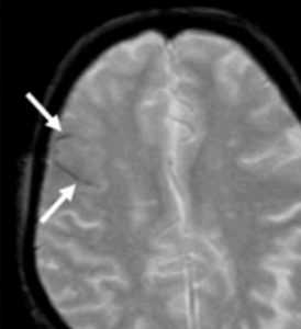
ARIA-E

On imaging, ARIA-E is most commonly observed on FLAIR or other T2-weighted sequences as hyperintensities in the parietal, occipital, and frontal lobes but has also been observed in the cerebellum and brainstem. The imaging features of ARIA-E are thought to represent edema in the gray and white matter as well as effusion of extravasated fluid in the sulcal space. The signal hyperintensities can be quite subtle in a single region, multifocal, or nearly pan-hemispheric.
Imaging characteristics of ARIA-E
Parenchymal vasogenic edema
Sulcal effusion
Swelling

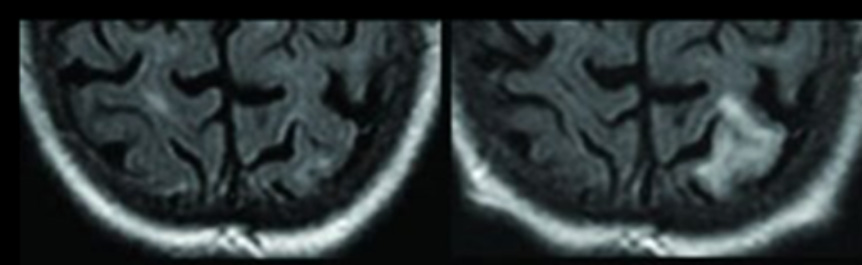
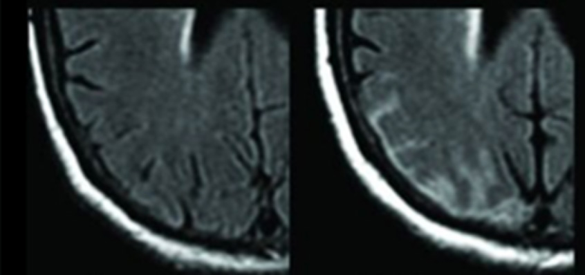
ARIA-E tends to be dose-dependent, with a greater risk of occurrence at higher doses of amyloid-targeting therapies. In addition, being an ApoE ε4 carrier has emerged as an important risk factor for treatment-related ARIA-E, potentially related to the associated higher parenchymal and vascular β-amyloid load. In these patients, anti-β-amyloid therapies could result in larger clearance, greater vascular permeability, and therefore easier extravasation of fluid (ARIA-E) and erythrocytes (ARIA-H).
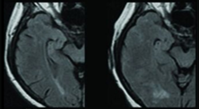
ARIA-H

ARIA-H results from iron deposits in the tissue in the form of hemosiderin. They are likely residuals of a small leakage of blood from a vessel into adjacent parenchymal tissue (microhemorrhages) or into adjacent subarachnoid space or peri-adventitial compartment (hemosiderosis).
Both microhemorrhages and hemosiderosis are detected on appropriately weighted T2 or T2* MRI sequences (such as GRE or SWI sequences). Without the sequences, the small amounts of hemosiderin cannot be detected because of the rephasing pulse gradients on T1, T2, and FLAIR imaging.
Microhemorrhages typically manifest as focal, round, very low-intensity (relative to adjacent brain) signals in the brain parenchyma. Hemosiderosis appears as curvilinear low intensities that lie adjacent to the brain surface.
Imaging characteristics of ARIA-H
Microhemorrhages
Superficial siderosis
The Alzheimer’s Association workgroup has recommended an ARIA-H cutoff of 4 microhemorrhages as an exclusion criterion for clinical trials of anti-Aβ monoclonal antibody treatment for AD. However, the risk of ARIA-H or ARIA-E due to baseline microhemorrhages has not been well-established and continues to be investigated. Further, the association is confounded by the variety of pathologic conditions that can include microhemorrhages on MRI and the increasing prevalence of baseline microhemorrhages with age.

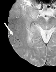
Size criteria have also been recommended, at cut-offs of ≤10 mm or ≤5 mm diameter. However, the technical features of image acquisition create challenges for consistently measuring the size of microhemorrhages. In addition, a “blooming” effect can occur: a phenomenon in which the apparent size of a microhemorrhage on MRI is larger than the size of the histologically defined hemosiderin deposit. This phenomenon is related to signal loss that extends spatially beyond the area of the deposit.

Therefore, in addition to meeting the label requirements, clinicians should consider genetic markers, contraindicated medical conditions, and the patient’s drug profile when prescribing Leqembi and other drugs in the same class. This holistic approach increases in importance when one takes into account the burden of infusions, varying efficacy, risk of side effects, and cost of treatment (at least US $25,000 a year). In the United States, the Centers for Medicare and Medicaid Services (CMS) has declined to cover Aduhelm under federal insurance plans unless the person was enrolled in a clinical trial, and Leqembi is currently covered under the same ruling. This leaves some patients paying the full cost out of pocket, increasing the importance of the best patient-drug match. Imaging and other biomarkers, even with their potential cost, will provide critical input into the most appropriate patients, their safety, and whether the drug is working as intended.
PET Amyloid Report from our cPET™ application (WIP: not cleared for clinical use)

Closely monitor efficacy and safety.

The heterogeneity of AD necessitates close monitoring of patients on amyloid-reducing DMDs, for optimal dosage and objective measurements of effectiveness. Endpoints related to cognitive decline can be subjective, vary between clinicians, and fail to capture changes in underlying pathology. Objective biomarkers such as imaging can be more accurate; for example, amyloid and tau PET scans were used to measure amyloid and tau accumulation, key endpoints in the phase 3 Leqembi trial.
Regarding safety, the most common reported side effects of Leqembi were infusion-related reactions, headache, and ARIA.
In clinical trials of amyloid-targeting therapies for AD, the incidence of ARIA has ranged from 0% to 42%. Compared with the 35% of patients who experienced ARIA with edema or effusions (ARIA-E) in clinical trials of Aduhelm, only 12.6% of patients receiving Leqembi experienced them (and 27% with donanemab). ARIA-E with Leqembi:
ARIA with microhemorrhages and superficial siderosis (ARIA-H) were experienced by 17.3% of the patients receiving Leqembi in the phase 3 clinical trial (compared with 9.0% in the placebo group).
The risk of ARIA depends on patient characteristics, the therapy’s mechanism of action, length of treatment, and dosage. Although most cases of ARIA in clinical trials (67-90%) of AD treatments were asymptomatic, headache, visual disturbance, dizziness, nausea and vomiting, and confusion have been reported. The most severe cases can require hospitalization, and the potential for a long-lasting impact on clinical course, including cognitive decline, and response to treatment is currently unknown. ARIA typically resolves after halting the medication, and the first few months represent the critical time period in which ARIA can occur.
Asymptomatic cases and treatment re-initiation will require close monitoring and the ability to detect subtle changes. Collecting and documenting a full clinical picture, including imaging, presence of symptoms, and potential risk factors, will help differentiate from other potential pathologies and to determine a diagnosis. Regarding imaging, the minimum standards for the specific MRI protocol proposed by the working group still appear to be valid:
Guidance provided by a recently convened expert panel in response to ARIA detected during clinical trials of Aduhelm included the acquisition of FLAIR, T2-weighted GRE or SWI, and DWI sequences within 1 year before treatment initiation and before the 5th, 7th, and 12th infusions during dose titration.
In addition, MRI should be performed whenever patients show any symptoms suggestive of ARIAs, and treatment should be discontinued for symptomatic patients. MRIs should then be obtained monthly to evaluate treatment reinitiation once ARIA-E has resolved or ARIA-H has stabilized. Otherwise, in the absence of symptoms, suspension of treatment is only recommended for radiologically severe or moderate ARIAs. However, close monitoring should occur to ensure no worsening of the ARIA condition.
The need for expertise in evaluating imaging for ARIA, as well as a standardized protocol, has been highlighted.
ARIA-H can be particularly challenging to accurately determine, and detection is dependent on the reader’s training and experience, features of image acquisition, image artifacts, regional signal loss, and potential confluence of multiple microhemorrhages. In addition, microhemorrhages can be particularly hard to discern in some brain areas such as paranasal sinuses and in the presence of dentures. There are times when a microhemorrhage might only become apparent when comparing MRIs from different time points for the same patient.
This is when validated algorithms can help to consistently and objectively detect, quantify, and monitor ARIA. For example, our cARIA™ application (still in development) has algorithms that support detection and quantification of ARIA-H (microhemorrhages and superficial siderosis) as well as ARIA-E.
Monitoring changes with MRI (ARIA-E and ARIA-H)
The results have been found to be accurate for assessing the severity of ARIA-H and ARIA-E.
Further, the results are presented in an easy-to-understand report for radiologists, clinicians, and patients.

Provide appropriate care across the patient pathway.

As genetic testing becomes more mainstream and celebrities like Chris Hemsworth shine the spotlight on genetic risk factors for AD, the public is likely to consider early access to DMDs an imperative when faced with a heightened genetic risk of cognitive decline. Aduhelm and Leqembi are the first drugs to enter this space, and with additional DMDs in development, neurologists and radiologists will be tasked with incorporating them into their AD armamentarium.
Each new DMD provides greater clarity about who would benefit, at the lowest risk, and helps prepare the health system, including establishing referral and diagnosis pathways as well as imaging and infusion infrastructure. Changes to reimbursement models for AD therapies could also improve uptake of new DMDs.
Given the challenging presentation and imaging characteristics of AD and ARIA, the right tools are needed to provide the best care for patients on DMDs. A technology-enabled imaging and diagnostic support tool can be an advantage for accurate patient management. Objective identification, measurement, and detection of changes in amyloid plaques or tau accumulation, micro-bleeds, and brain swelling can differentiate neurology and radiology practices in terms of the care provided and patient safety. Detailed, clear reporting can ease concerns from physicians, patients, and caregivers alike concerning the benefits and risks of new drugs.

Will you be attending ECR 2023?

Stop by our booth, EXPO X1 in the Artificial Intelligence Exhibition (AIX) area AI-36, for a demonstration of our artificial intelligence (AI)–enabled software solution that was designed to support entire patient care pathways, including early detection, differentiation of diagnosis, treatment planning, and monitoring.