
cNeuro® cPET and cDAT
cPET provides precise quantification of amyloid and FDG PET brain scans with comparison to reference data from healthy controls.
cDAT provides precise quantification of DAT SPECT images with comparison to reference data from healthy controls.
cNeuro® cPET and cDAT, integrates with PACS, and results can be viewed in PACS or using the cNeuro browser-based viewer.
Nuclear medicine physicians benefit from support to in detecting subtle changes and increased confidence in reading borderline cases.
Capabilities of cPET
Two modes of operation for precise quantification. Supports PET-only and PET-MRI workflows; if the MRI is available, PET findings can be combined with information about atrophy and lesions from MRI. PET-only utilizes a novel multi-atlas image registration method.
Fully automated, reliable, reproducible analysis of amyloid and FDG brain PET scans:
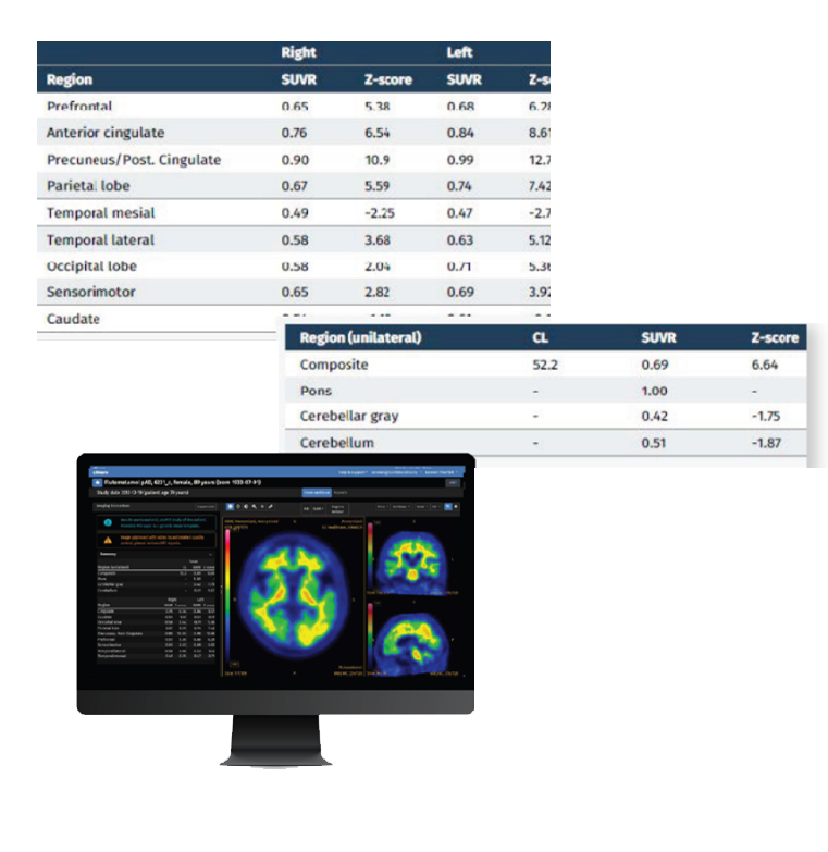
- Regional standard uptake value ratios (SUVRs) are computed for cortical and sub-cortical structures
- Regional and image-based Z-scores computed based on comparison with tracer-specific reference data
- PET data superimposed on MRI (patient’s MRI or MRI template) allow findings to be correlated to patient anatomy
Capabilities of cDAT
Fully automated analysis of DAT SPECT images:
- Quantified features are compared to age-matched healthy controls and z-scores are computed. The Normal Database is based on data from the Parkinson’s Progression Markers Initiative (PPMI)
- Computes striatal binding ratios of the caudate and putamen VOIs, compared to a background region, as well as putamen/caudate ratios and asymmetry indexes
- Orients the image automatically for optimal visual assessment and removal of head-tilt artifacts

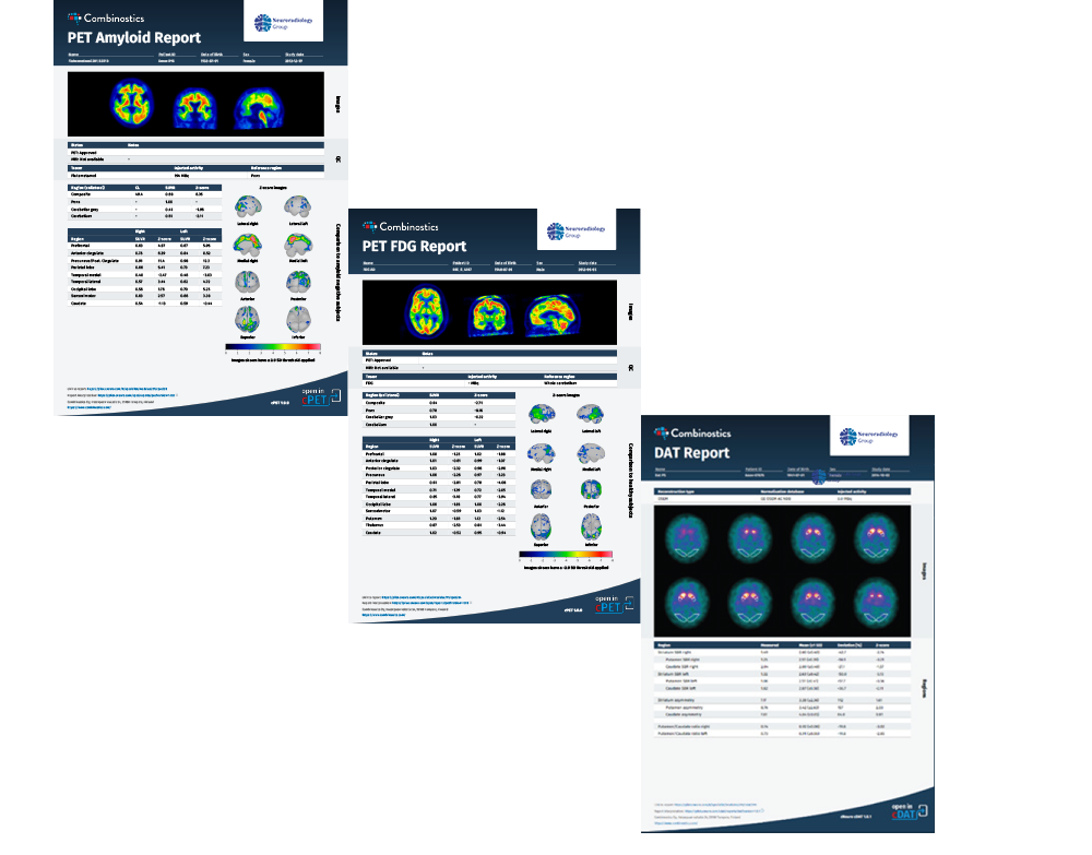
Improve collaboration and referrals with clinically focused reports
Improve collaboration, provide greater value, reduce image-related queries, and drive more referrals through streamlined, high-quality, consistent communication with referring clinicians.
Our cPET reports for amyloid and FDG brain scans and cDAT reports for DAT SPECT provide information about the patient’s status and facilitate patient management as well as communication between nuclear medicine physicians, neurologists, patients, and their caregivers.
Regulatory Compliance
cPET: CE marked, FDA 510(k) cleared
cDAT: CE marked, FDA 510(k) cleared
Security
- All data transfer uses SSL encryption
- Stored data are anonymized and encrypted
- More information is available in a separate security statement.
cNeuro cPET Indications for Use
cNeuro cPET aids physicians in the evaluation of patient pathologies via assessment and quantification of PET brain scans.
The software aids in the assessment of human brain PET scans enabling automated analysis through quantification of tracer uptake and comparison with the corresponding tracer uptake in normal subjects. The resulting quantification is presented using volumes of interest and voxel-based maps of the brain. cNeuro cPET allows the user to generate information regarding relative changes in PET-FDG glucose metabolism.
cNeuro cPET additionally allows the user to generate information regarding relative changes in PET brain amyloid load between a subject’s images and a normal database, which may be the result of brain neurodegeneration.
PET co-registration and fusion display capabilities with MRI allow PET findings to be related to brain anatomy.
cNeuro cPET aids physicians in the image interpretation of PET studies conducted on patients being evaluated for cognitive impairment, or other causes of cognitive decline.
cNeuro cDAT Indications for Use
cNeuro cDAT is intended for use by Nuclear Medicine or Radiology practitioners and referring physicians for display, processing, and reporting of Nuclear Medicine Imaging data.
cNeuro cDAT enables visual evaluation and quantification of 123 Ioflupane (DaTscan™/DaTSCAN™) images. The software enables automated quantification of tracer uptake and comparison with the corresponding tracer uptake in healthy subjects as provided by normal population databases of 123 Ioflupane (DaTscan™/DaTSCAN™) images. cNeuro cDAT assists in detection of loss of functional dopaminergic neuron terminals in the striatum, which is correlated with diseases associated with dopaminergic deficiencies, such as Parkinson disease or Dementia with Lewy Bodies (DLB).

