BLOG

The ideal state of neuroradiology according to Dr. Kevin Berger? More objective data and more integrations in software-driven imaging quantification

One benefit of expanding our reach in radiology practices globally is the opportunity to work with pioneers in the field, hearing about their experiences and learning about how we’re meeting specific pain points with our products and what we can do to make our applications even more effective for our end users. We recently spoke with Kevin Berger, MD, a radiologist specializing in diagnostic radiology and neuroradiology at Chesapeake Medical Imaging. He has a long history of contributing to the development of imaging modalities such as PET and MRI, including as a consultant to the U.S. National Institutes of Health, as well as quantification tools. He recently started using our cMRI™ application and is excited about cMRI’s approach to quantifying patient images.

Read on to learn more about this as well as Dr. Berger’s history in neuroradiology and how he’s seen imaging quantification progress over the years.

What sparked Dr. Berger’s interest in neuroradiology?

1989 — during an undergraduate honors program at the University of Michigan, Dr. Berger had the opportunity to conduct research using PET functional imaging to assess blood flow in the brain. This initiated an interest in neuroradiology that continued in his diagnostic radiology residence and neuroradiology fellowship at Mallinckrodt Institute of Radiology at Washington University in St. Louis. During his academic career at Michigan State University, he published findings related to multi-modal imaging of Alzheimer’s disease and mild cognitive impairment, while in his clinical practice, Dr. Berger has used the range of imaging modalities (e.g., PET, CT, MRI) as well as imaging quantification tools.

He was instrumental in developing a software program using PET to diagnose dementias, which later was licensed and became available commercially. Since then, he’s advised on the development of other similar programs using PET or MRI, has contributed as the lead clinical site for the validation of imaging software programs, and has assisted companies with obtaining regulatory clearance for their software programs to assess neurodegenerative diseases. As he said, he knows firsthand that “for imaging quantification software, the timeline from research application to clinical use can be long.”

Have quantitative imaging programs become more accepted by radiologists?

Similarly, acceptance of quantitative imaging programs by radiologists has also taken some time, but Dr. Berger has observed that the benefits of objective imaging measurements for improved performance and enhanced validity are increasingly being recognized by his radiology colleagues as well as referring physicians. Having quantitative analysis to guide the interpretation can increase confidence in the assessments.
Dr. Berger finds that his referring physicians have greater confidence in the results when he explains the software programs are helping him help them — through objective, quantitative analysis to guide his interpretation and a computer algorithm that validates what he visually observes. As he said, “physician confidence in the outputs increases when they hear someone with 30 years of experience say that validation of my findings by a computer algorithm helps me help them with a more confident assessment.”
Although most radiology practices are currently using some form of quantitative software, adoption will also likely accelerate as insurance companies increasingly require quantitative analysis for approval of payment of more advanced imaging when required.

When can quantitative imaging assessments have the greatest impact?

For most neurological conditions, subjective visual assessment could be sufficient for an assessment at a single point in time, surmised Dr. Berger. However, for dementias in particular, a single quantitative assessment can be useful to make a specific diagnosis, by providing prognostic information in the form of brain morphometric patterns. And with the introduction of disease-modifying drugs (DMDs) such as ADUHELM and LEQEMBI for Alzheimer’s disease, being able to make a specific diagnosis with a sufficient level of probability based on objective, quantifiable measures of disease is of paramount importance.
For other diseases such as multiple sclerosis, quantitative, objective imaging assessment is already improving the ability to monitor longitudinal changes in lesion count and burden. After all, subtle changes in disease measures are better detected and quantified by a computer than the human eye, increasing the validity of the assessment and better informing disease management and medication decisions.

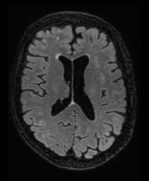
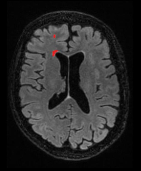
What role does cMRI have in Dr. Berger’s practice?

In Dr. Berger’s practice, cMRI has been used for approximately 6 months now and is contributing to their ability to provide their referring physicians with accurate, reliable, and objective quantification of brain imaging. It was relatively simple to integrate into their workflows: once the patient data set is uploaded to the cloud-based app (either automated using the gateway or manually by a technologist), the resulting data and analysis are transferred back to their PACS. It’s largely offline and automated and provides a considerable amount of data.
When asked about what makes cMRI stand out from other quantification tools, he’s excited about cMRI’s approach to register the patient data, which has been on his wish list for the past 10 years. Because it compares a systematically selected set of atlases against the patient image within the patient space, cMRI can generate more accurate results and reduce the likelihood of false positive findings. In this approach, each atlas is registered non-rigidly (“stretched”) to the patient image to estimate the anatomical correspondence of each segment between the atlas and the patient image.
In comparison, other tools compare the patient image against a standardized atlas space. In this approach, partial volume segmentation is more likely to occur. When this happens, the image intensity may not represent any tissue class, which can contaminate the data and result in less-than-desired accuracy of the final data. Ventricle size is one area that comes to mind for this problem. Because ventricles easily overlap grey matter space using this standardized space method, the measurement of ventricle size is problematic.
In his practice, he finds that the enhanced differentiation between conditions based on accurate quantification is a valuable benefit of cMRI. For example, in this 63-year-old patient with worsening memory, there is a very low burden of disease, which is challenging to quantify visually (left). However, with cMRI, the amount of white matter hyperintensities (WMH) is more apparent.
Axial FLAIR image
WMH identified by cMRI
The Dementia Report generated by cMRI provides additional information for the assessment of this patient: significant global atrophy but not significant hippocampal atrophy on the plots, when compared with controls without dementia (indicated by the red lines).
The anterior versus posterior score is -3.7, which ranks in the first percentile and shows differential atrophy in the anterior versus posterior areas of the cortex. This is predictive of frontotemporal dementia (FTD), supported by the 50% likelihood of FTD reported by cMRI (compared with 20% likelihood each of Alzheimer’s disease or cognitively normal). Dr. Berger was quick to point out that the age-matching and consideration of disease prevalence by cMRI are important for determining the probability of a diagnosis. For example, some diseases are less prevalent for different age groups, such as FTD in older age. Therefore, the relative atrophy observed in this patient might not have the same significance for an older patient.
According to Dr. Berger, cMRI offers the superior approach that provides more accurate, reliable, and valid data that are immediately usable. This is why cMRI’s reliability and validity were not at top of mind when asked about how he would improve the application. Instead, his response was more process-oriented, with the aim of improving reimbursement. He is interested in receiving only the relevant data for the patient’s specific clinical scenario: His future state would have the radiologist inserted throughout the process to provide quality control and specify which volume areas to assess and return to PACS, before reviewing quantitative analysis results.

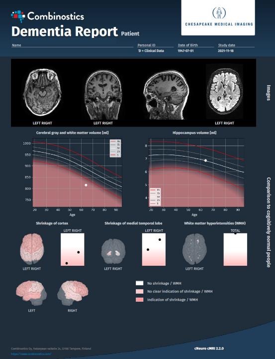
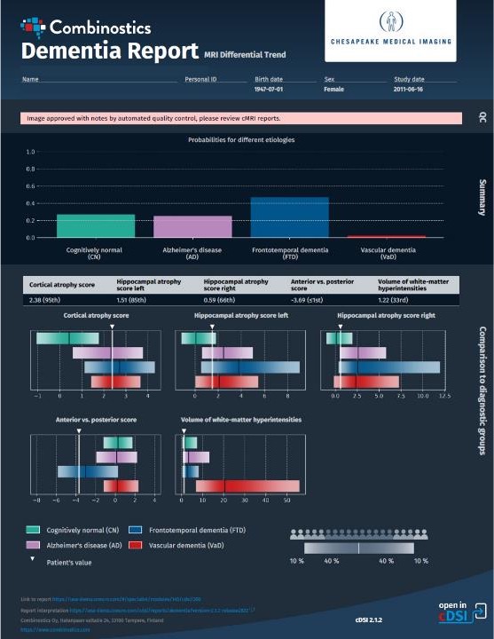
The end goal is a more holistic approach to patient assessment.

More, more, more — objective assessments, radiologist involvement, artificial intelligence–driven assessments, and integration of clinical data. According to Dr. Berger, “a platform that provides a combination of imaging modalities and clinical neuropsychometric data will give us more specific and better answers than any parceled approach alone.” And, cMRI is well equipped to handle more and fit within such a platform to enhance patient care.

We’re looking forward to continuing to work with Dr. Berger and benefit from his insights on radiology practice, imaging software, and more. If you’d like to learn more about cMRI, contact us for a discussion or a demo, or take advantage of our free 10-day trial of cMRI on your own patient cases.