BLOG

Slicing through the fog of neurodegeneration with cutting-edge tools: feedback from ASNR22

By: Peter Ngum, Clinical Application Specialist, and Greg Kingston, Chief Marketing Officer (CMO)
2022 seems to be the year of the conference, and we’re loving every minute of it. From advances in the treatment of multiple sclerosis (MS) at ACTRIMS 2022 to broader discussions about neurology at AAN 2022, our team has had the chance to meet up with old and new colleagues and learn about innovative solutions across the field. We’ve just returned from the Annual Meeting of the American Society of Neuroradiology (ASNR22) in New York City. In its 60th year, the theme of ASNR22 was “Reconnecting the Global Neuroradiology Community,” and from the vantage point of our booth in the exhibit hall, it succeeded at stimulating great conversations, some of which we describe here.

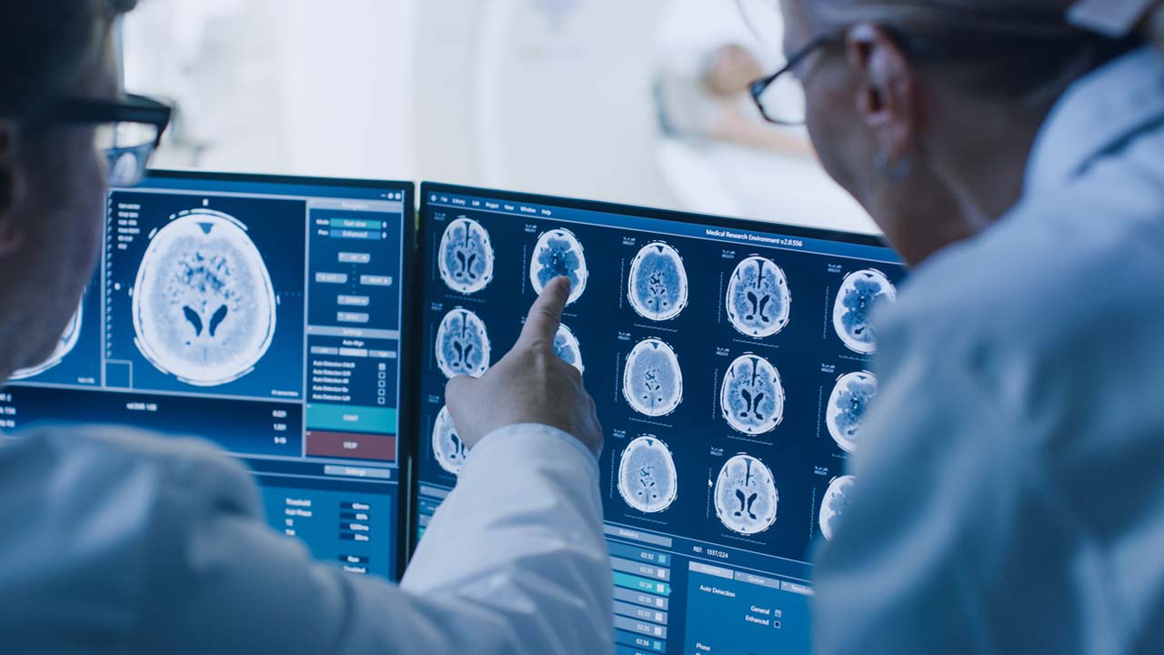
Clinicians value tools that can help them confidently diagnose dementias.

We were particularly excited to discuss and hear feedback on our new Dementia Differential Analysis report, which we recently announced and presented in a session at the conference. Our team has worked hard to generate visualizations based on comparisons of patient MRI biomarkers with a historical database of people with normal cognition and 2000+ patients with various types of dementia.

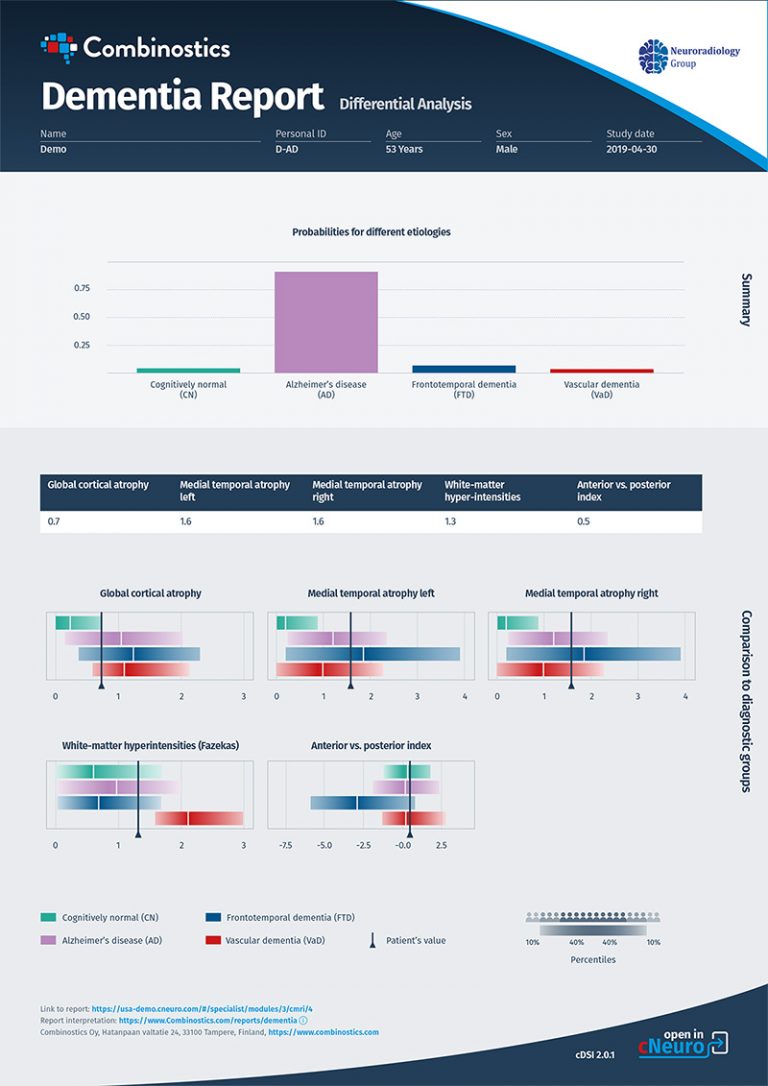
The report is based on MRI data only, using our cDSI™ application’s proprietary machine learning method that measures the similarity of the following disease-specific imaging biomarker values from the patient to those of different diagnostic groups:
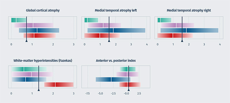
Feedback at the booth and during the presentation was positive, reflecting our belief that this report could make a difference in the diagnosis and management of patients with symptoms of dementia. Many different pathologies present with dementia-related symptoms, and determining the underlying etiology is often based on assessments of cognitive function combined with subjective visual evaluations of patterns of atrophy on imaging — or by merely excluding easily identifiable causes of cognitive impairment such as tumors or hemorrhages.
Yet, it is important to be able to give a confident diagnosis between normal cognition, Alzheimer’s disease (AD), frontotemporal dementia (FTD), and vascular dementia (VaD) for disease management, especially as disease-modifying therapies (DMT) become available. However, neuroradiologists tend to only rule dementia in or out rather than provide a specific dementia diagnosis when reporting to the referring clinician, unless they received a specific request for diagnostic options based on a degree of certainty. In other words, the referring clinician often guides the reporting.
It was encouraging to hear senior neuroradiologists see the value of using the report to help them understand the split between the different types of dementia and give confidence in the diagnostic decision. The visualizations were perceived as a great communication tool to share with referring clinicians. The probabilities graph was especially impactful — they show the distributions based on the database of patients and where the current patient fits on the distribution, guiding the diagnosis.

Applying artificial intelligence to neuroradiology requires a multidisciplinary approach.

The potential of artificial intelligence (AI) applications to change the field of neuroradiology was featured in AI-specific workshops and tracks. The presentations highlighted the need for a multidisciplinary approach to AI system development — including neurology, physics, statistics, and machine learning expertise — before it can be applied in a clinical setting. And there’s a wide range of disease applications, from brain tumors to neurodegeneration to pediatric settings.

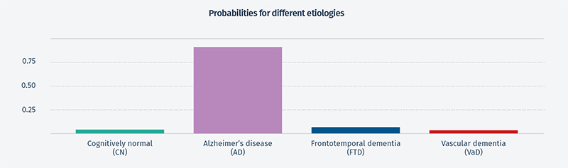
Although clinicians don’t need to understand the development process or the science and technology behind the outputs, they do want to know the system has been validated. This was a question of interest regarding our new report from neuroradiologists at our booth. Collaborating with clinicians from memory clinics, we have been able to show 76% accuracy in separating AD, FTD, VaD, and normal cognition using MRI data in cDSI, which is much higher than other methods. The remaining 24% of cases comprised mixed dementia diagnoses and pre-clinical disease, among others. For the 50% of patients with the highest probability, the accuracy increased to 88%. When additional patient variables were added to cDSI, such as cognitive test results, cerebrospinal fluid test results, and genetic test results, the diagnostic accuracy increased to 88% for all patients and 99% for the 50% of patients with the highest probability.

We’re looking forward to next year.

We had meaningful conversations during ASNR22 with a number of neuroradiologists whose specialties align with our focus on neurodegenerative diseases, including dementias, MS, and traumatic brain injuries and who are interested in the latest developments to improve their practice. The strong audience, combined with the academic nature of the conference, contributed to a positive, high-value experience, and when asked if we’ll attend again next year, our answer is “Absolutely.”

Until then, schedule a meeting to chat or stop by and see us at booth Nord 39 at the 103rd German X-Ray Congress.